Eagleview Eye Clinic

Archives August 2025

SCLERA

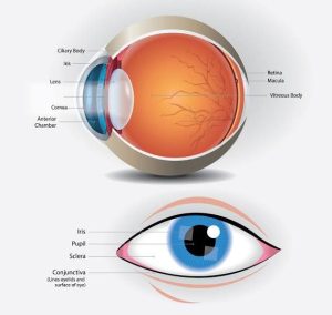
The sclera, also known as the white of the eye, is the opaque, fibrous, and protective outer layer of the eyeball. It provides structure, protection, and shape to the eye.

Key characteristics:

  1. Composition: The sclera is made of dense, collagenous connective tissue.
  2. Location: It covers the posterior five-sixths of the eyeball, with the cornea covering the anterior one-sixth.
  3. Function: The sclera:
    • Provides protection to the intraocular contents
    • Maintains the shape of the eyeball
    • Serves as an attachment site for the extraocular muscles that control eye movement
  4. Appearance: The sclera is typically white or off-white in color, but it can appear differently in certain conditions.

Importance: The sclera plays a vital role in maintaining the integrity and function of the eye, and its health is essential for preserving vision.
BLEB

A bleb is a blister-like structure that forms on the surface of the eye, typically as a result of a surgical procedure, such as trabeculectomy. In the context of glaucoma surgery, a bleb is a fluid-filled reservoir that forms under the conjunctiva, allowing excess fluid to drain from the eye and reducing intraocular pressure.

Characteristics of a bleb:

  • Fluid-filled
  • Located under the conjunctiva
  • Can be diffuse or localized
  • May be elevated or flat

The presence and characteristics of a bleb can indicate the success of the surgery and the effectiveness of IOP reduction.

TRABECULECTOMY

Trabeculectomy is a surgical procedure used to treat glaucoma by creating a new drainage pathway for fluid to leave the eye, reducing intraocular pressure (IOP). The goal is to:

  • Lower IOP to prevent further damage to the optic nerve
  • Preserve vision
  • Slow disease progression

During the procedure:

  • A small section of tissue is removed from the eye
  • A new drainage channel is created
  • A bleb (fluid reservoir) forms under the conjunctiva

Trabeculectomy is often considered for patients with:

  • Advanced glaucoma
  • Uncontrolled IOP despite medication or laser treatment

Post-operative care includes:

  • Monitoring for complications (e.g., infection, scarring)
  • Managing IOP and inflammation
  • Follow-up appointments for recovery assessment.

NEUROPARALYTIC KERATITIS

Neuroparalytic keratitis is a condition where the cornea becomes inflamed or damaged due to nerve dysfunction or paralysis, often resulting from:

  • Facial nerve palsy (e.g., Bell’s palsy)
  • Trigeminal nerve damage
  • Trauma
  • Stroke
  • Neurological disorders (e.g., multiple sclerosis)

This condition can lead to:

  • Decreased corneal sensation
  • Reduced blinking
  • Exposure keratopathy
  • Increased risk of corneal ulcers

Symptoms may include:

  • Eye pain
  • Redness
  • Blurred vision
  • Dryness

Treatment aims to:

  • Protect the cornea
  • Promote healing
  • Manage symptoms

Options may include:

  • Lubricating eye drops
  • Protective eyewear
  • Tarsorrhaphy (surgical eyelid closure)
  • Other supportive measures.
ORBITAL TUMOUR REMOVAL

Orbital tumor removal is a surgical procedure to remove abnormal growths or tumors within the orbit (the bony cavity surrounding the eye). The goal is to:

  • Eliminate the tumor
  • Preserve vision and eye function
  • Improve appearance
  • Relieve symptoms (e.g., pain, double vision)

Types of orbital tumors:

  • Benign (non-cancerous): e.g., hemangiomas, neurofibromas
  • Malignant (cancerous): e.g., lymphoma, rhabdomyosarcoma

Surgical approaches:

  • Anterior orbitotomy: For tumors located in the front part of the orbit.
  • Lateral orbitotomy: For tumors located on the side of the orbit.
  • Cranio-orbital approach: For tumors extending into the cranial cavity.

Techniques:

  • Open surgery: Traditional approach with incisions.
  • Endoscopic surgery: Minimally invasive approach using small incisions and a camera.

Post-operative care:

  • Monitoring for complications (e.g., bleeding, infection)
  • Managing pain and swelling
  • Follow-up appointments for recovery assessment

The specific approach and technique depend on the tumor’s location, size, and type, as well as the patient’s overall health.