Eagleview Eye Clinic

Archives 2025

KERATOPLASTY

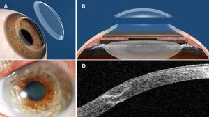
Keratoplasty or cornea grafting is a surgical procedure where a damaged cornea is replaced with a donated cornea tissue.

The cornea is the transparent outermost tissue of the eye over lapping the iris. Most people refer to the cornea as the black of the eye.

Conditions that can necessitate a cornea grafting include keratoconus, cornea decompensation, iridocornea endothelium syndrome, et.c

If the whole cornea is been replaced, then it is known as penetrating keratoplasty and if a part of the cornea is been replaced then it’s known as lamellar keratoplasty .
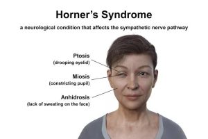
HORNER’S SYNDROME

Horner’s syndrome is a neurological disorder that occurs when the oculosympathetic nerve is damaged.

It can be caused by a tumour, stroke or damage to the middle ear..

When the sympathetic nerves that connects the brain stem to the eye is damaged a triad of ptosis ( Droopy lids), miosis (Constricted pupils) and anhidrosis ( lack of sweat) occurs.

Although this condition is similar to Bell’s palsy, it is different from Bell’s palsy.

Treatment basically involves treating the underlying cause.

PIGMENT DISPERSION SYNDROME

This is a condition whereby the pigments from the iris sloughs off and blocks the trabecular meshworks leading a buildup of aqueous humor and consequently increased IOP and a particular type of glaucoma known as pigmentary Glaucoma.

The cause of this condition is unknown…

This condition most of the time is usually asymptomatic until a large proportion of the optic Nerve have been damaged during to Glaucoma. Another sign of the pigment dispersion syndrome is the krekunberg spindle which is a vertical pigmentation on the endothelium of the cornea caused by iris fragments.

Treatment usually involves managing the glaucoma that results from this condition.

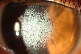
CORNEA DYSTROPHIES

This is genetic condition whereby abnormal materials are deposited  between the layers of the cornea ( the outer black part of the eye).

This usually results in severe reduction in vision..

Treatment is strictly keratoplasty either penetrative or lamellar, depending on the layer of the cornea that is affected.

EPIRETINAL MEMBRANE

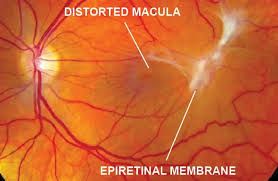
Epiretinal membrane also known as cellophane maculopathy or macular pucker is an avascular, Semi translucent, fibrocellular formed over the Retinal surface.

This membrane is caused by changes in vitreous integrity, retinal detachment et.c

This condition have been known to cause painless loss of vision.

Most of the time epiretinal membrane is asymptomatic but it can result in diplopia, metamorphopsia.

Treatment options involves surgical intervention.

SYNCHISIS SCINTILLANS

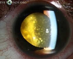
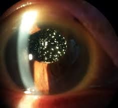
SYNCHISIS scintillans, also known as cholesterol bulbi are yellowish deposits of fine cholesterol crystals in the vitreous body of the eye.

Usually SYNCHISIS scintillans is usually asymptomatic and is seen by accident.

It is most times usually as a result of degenerative changes of the vitreous due to age.

Treatment is usually by surgical procedure.

Difference between SYNCHISIS scintillans and asteroid hyalosis, is that while SYNCHISIS scintillans floats within the vitreous, asteroid hyalosis is fixed.

ANIRIDIA

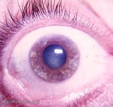
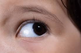
ANIRIDIA is simply the absence of a muscular structure called the Iris.

The iris forms the pupil which controls how light enters the eyes. The iris is also responsible for eye color. So incases of Aniridia, the eyes just appear black.

This condition can be congenital, affecting both eyes or traumatic due to a penetrating injury.

For some people Aniridia can result in a significant loss of vision, while for  others visual reduction mild.

Use of contact lenses can help control light and improve vision. Use of photochromic glasses or sunshades can also help to significantly improve vision.

ASTEROID HYALOSIS

Asteroid HYALOSIS are yellowish glittering particles deposits on the vitreous body caused by age related vitreous degeneration.

This condition is usually age related and does not affect vision in any way.

Most of times, the patients does not even notice any sign or symptom but a common symptom of this condition is floaters ( moving objects in the field of view).

If the symptom becomes  discomforting, surgery is usually the way out for the patient.

HERPES ZOSTER OPHTHALMICUS

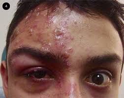
This condition is simply shingles that involves the eyes and surrounding areas.

This condition is caused by reactivation of the varicella zoster virus in the trigeminal ganglion supplying the ophthalmic division of the trigeminal nerve.

Signs and symptoms includes characteristic rash on one side of the face, pain, itching, blurring vision and on rare cases keratitis.

Immunocompromised patients, patients who have had chicken pox in the past are at risk of contracting herpes zoster OPHTHALMICUS.

Treatment includes the use of antiviral tablets, and antiviral ointment, and anti- inflammatory drugs.

OPTIC NEURITIS

Optic neuritis is an inflammation of the optic nerves. The optic nerves are connections between the eye and the brain responsible for transmission of visual impulses between the two organs

This condition is caused by autoimmune ailments like multiple sclerosis, myasthenia gravis and viral infections like shingles , chicken pox et.c

This condition is sight threatening, especially if not treated early.

Signs and symptoms includes pain on eye movement, reduced vision, defective colour vision, eye pain.

Treatment involves treating underlying conditions, and the use of corticosteroids.