Eagleview Eye Clinic

Tag eye care professionals

LAYERS OF THE IRIS

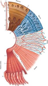
The iris consists of several layers:

  1. Anterior border layer: The front layer of the iris, consisting of fibroblasts and melanocytes.
  2. Stroma: The middle layer, composed of connective tissue, blood vessels, and melanocytes that give the iris its color.
  3. Anterior epithelium (Anterior pigmented epithelium): A layer of pigmented cells.
  4. Posterior epithelium (Posterior pigmented epithelium): A layer of densely pigmented cells that block light from entering the eye except through the pupil.

These layers work together to control the amount of light entering the eye by adjusting the size of the pupil and give the iris its color and structure.
LAYERS OF THE CORNEA

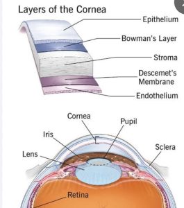
The cornea, the transparent outer layer of the eye, consists of five distinct layers:

  1. Epithelium: The outermost layer, composed of stratified squamous epithelial cells, providing a barrier against external factors.
  2. Bowman’s layer: A dense, acellular layer of collagen fibers beneath the epithelium, providing strength and structure.
  3. Stroma: The thickest layer, composed of collagen fibers and keratocytes, making up about 90% of the cornea’s thickness.
  4. Descemet’s membrane: A thin, porous layer of collagen fibers produced by endothelial cells.
  5. Endothelium: The innermost layer, composed of a single layer of endothelial cells, responsible for maintaining corneal clarity by regulating fluid and electrolyte balance.

These layers work together to maintain the cornea’s transparency, structure, and function.

CHOROID

The choroid is a complex vascular layer in the eye, situated between the sclera and retina. It consists of blood vessels, melanocytes, and fibroblasts.

Key characteristics:

  1. Blood supply: The choroid is richly vascularized, supplying the retina with oxygen and nutrients.
  2. Location: It’s situated between the sclera (outer layer) and retina (inner layer).
  3. Melanin content: The choroid contains melanin, which helps absorb excess light.

Functions:

  1. Nutrient supply: Provides the retina with essential nutrients and oxygen.
  2. Waste removal: Helps remove waste products from the retina.
  3. Light absorption: The melanin in the choroid absorbs excess light, reducing scatter and improving vision.

The choroid plays a vital role in maintaining the health and function of the retina, supporting clear vision and overall eye health.

VITREOUS BODY

The vitreous body, or vitreous humor, is a clear, gel-like substance that fills the space between the lens and the retina in the eye. It:

  1. Maintains eye shape: Gives the eye its spherical shape.
  2. Supports retina: Helps keep the retina in place.
  3. Transmits light: Allows light to pass through to the retina.

The vitreous body is composed of:

  1. Water: About 99% water.
  2. Collagen fibers: Provides structure and support.
  3. Hyaluronic acid: Helps maintain its gel-like consistency.

As we age, the vitreous body can undergo changes, such as:

  1. Vitreous detachment: Separation from the retina.
  2. Floaters: Small particles that cast shadows on the retina.

Issues with the vitreous body can lead to conditions like retinal detachment or vision problems.

ZONULE FIBRES

Zonular fibers, also known as suspensory ligaments or zonules of Zinn, are tiny, thread-like structures in the eye. They:

  1. Connect the lens to the ciliary body: Zonular fibers attach the crystalline lens to the ciliary body.
  2. Suspend the lens: They hold the lens in place, allowing it to change shape for focusing.
  3. Transmit forces: Zonular fibers transmit forces from the ciliary muscles to the lens, enabling accommodation.

Zonular fibers play a crucial role in:

  1. Lens accommodation: They help change the lens shape for near or far vision.
  2. Maintaining lens position: They keep the lens centered and stable.

Dysfunction or damage to zonular fibers can lead to vision problems, such as:

  1. Lens subluxation or dislocation
  2. Vision disturbances

Zonular fibers are an essential part of the eye’s anatomy, supporting the lens and enabling clear vision.

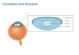
CRYSTALLINE LENS

The crystalline lens is a clear, flexible structure behind the iris and pupil in the eye. It plays a crucial role in focusing light onto the retina, enabling clear vision.

Key functions:

  1. Focusing light: The lens changes shape to focus light on the retina, allowing us to see objects clearly at varying distances.
  2. Accommodation: The lens adjusts its shape to focus on near or far objects, a process controlled by the ciliary muscles.

Characteristics:

  1. Transparency: The lens is clear, allowing light to pass through.
  2. Flexibility: The lens changes shape to focus light.
  3. Protein composition: The lens is made up of proteins and water.

Common issues:

  1. Cataracts: Clouding of the lens, affecting vision.
  2. Presbyopia: Age-related loss of near vision due to reduced lens flexibility.

The crystalline lens is a vital part of the eye’s anatomy, and its proper function is essential for clear vision.

CILIARY BODY

The ciliary body is a ring-shaped structure in the eye, located behind the iris. It’s a crucial part of the eye’s anatomy, responsible for:

  1. Aqueous humor production: The ciliary body produces the clear fluid (aqueous humor) that fills the anterior and posterior chambers of the eye.
  2. Lens accommodation: The ciliary muscles control the shape of the lens, allowing it to focus on near or far objects.
  3. Lens suspension: The ciliary body is connected to the lens via zonular fibers, which suspend the lens in place.

The ciliary body consists of two main parts:

  1. Ciliary muscles: These muscles contract and relax to change the lens shape for focusing.
  2. Ciliary processes: These are finger-like extensions that produce aqueous humor.

The ciliary body plays a vital role in maintaining the eye’s health and function, and any issues with it can lead to vision problems or eye diseases.

IRIS

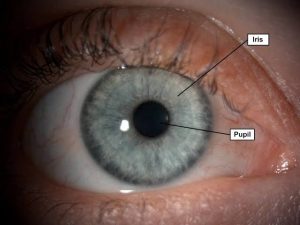
The iris is the colored, circular structure in the eye, surrounding the pupil. It’s a complex and highly specialized tissue that plays a crucial role in regulating light entry and supporting overall eye health.

Key characteristics:

  1. Color: The iris gets its color from melanin pigments, resulting in a wide range of colors and shades.
  2. Structure: The iris consists of two main layers:
    • Stroma (front layer): Contains collagen fibers, blood vessels, and melanocytes.
    • Epithelium (back layer): Contains pigment cells and muscles that control pupil size.
  3. Muscles: The iris contains two types of muscles:
    • Sphincter pupillae: Constricts the pupil in bright light or during near vision.
    • Dilator pupillae: Dilates the pupil in low light or during emotional arousal.
  4. Function: The iris:
    • Regulates light entry into the eye by adjusting pupil size.
    • Helps maintain optimal vision and protects the retina from excessive light exposure.
  5. Uniqueness: Each individual’s iris has unique patterns, colors, and textures, making it useful for identification purposes.

Importance: The iris plays a vital role in controlling light exposure to the retina, supporting optimal vision, and contributing to overall eye health.

PUPIL

The pupil is the circular opening in the center of the iris, the colored part of the eye. It plays a crucial role in regulating the amount of light that enters the eye.

Key characteristics:

  1. Location: The pupil is located in the center of the iris.
  2. Function: The pupil:
    • Regulates light entry into the eye
    • Adjusts size to control light intensity
    • Helps maintain optimal vision
  3. Size adjustment: The pupil:
    • Constricts (gets smaller) in response to:
      • Bright light
      • Near vision
      • Certain medications
    • Dilates (gets larger) in response to:
      • Low light
      • Emotional arousal
      • Certain medications or substances
  4. Control: The pupil’s size is controlled by the autonomic nervous system, specifically the:
    • Parasympathetic nervous system (constriction)
    • Sympathetic nervous system (dilation)

Importance: The pupil’s ability to regulate light entry is essential for maintaining optimal vision, protecting the retina, and responding to changes in lighting conditions.

CONJUNCTIVA

The conjunctiva is a thin, transparent, mucous membrane that covers the anterior (front) part of the eye and the inside of the eyelids. It plays a crucial role in maintaining eye health and function.

Key characteristics:

  1. Location: The conjunctiva covers:
    • The sclera (white part of the eye)
    • The inside of the upper and lower eyelids (palpebral conjunctiva)
    • The fornices (areas where the eyelids meet the eye)
  2. Function: The conjunctiva:
    • Produces mucus to lubricate the eye and facilitate smooth movement
    • Helps protect the eye from infection and foreign particles
    • Aids in maintaining the health of the cornea
  3. Structure: The conjunctiva consists of:
    • Epithelial cells
    • Substantia propria (a layer of connective tissue)
  4. Importance: The conjunctiva is essential for:
    • Maintaining eye comfort and moisture
    • Preventing infection and inflammation
    • Supporting the overall health of the eye

Common conditions: Conjunctivitis (pink eye), allergies, and dry eye syndrome can affect the conjunctiva, causing symptoms such as redness, itching, discharge, and discomfort.