Eagleview Eye Clinic

Archives January 2025

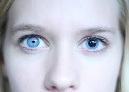
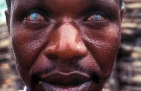
ONCHOCERCIASIS

Onchocerciasis or river blindness is caused by the parasitic worm known as onchocercha vovulus.

This condition occurs when an individual is bitten by black flies hence injecting the larvae of these worms into the host.

The larvae takes about 12 – 18 months before attaining maturity stage, they then move into the skin and blood stream wrecking havoc in their wake.

This condition is transmissible, as larvae of these worms are found underneath the skin of infected people,  hence if an infected person is bitten by black fly, the fly picks up the larvae from infected persons and transmit them to non infected persons when bitten.

People who dwell in riverine areas are more prone to this condition, hence the reason it is called river blindness.

Signs and symptoms includes eye pain, cloudy cornea, itching skin, swollen skin, et.c.

Treatment involves the use of ivermectin at a  6 month interval. Although ivermectin does not kill adult filarial worms.
LOIASIS

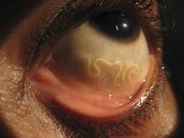
Loa loa filariasis or LOIASIS is caused by the loa loa nematode.

This loa loa is known as the African eye worm as it basically localizes in the conjunctiva.

It is caused by a bite from the deer flies or horse flies. When this parasitic flies bites it’s hosts, the larvae of this worms settle in blood and lymphatic system, lungs or underneath the skin. they can stay for as long as 15 – 17 years in the host body.

This condition is most common in people residing in rainforest Africa.

Signs and symptoms includes visible worm meandering within the conjunctiva, swollen skin, itching, et.c.

This condition is not sight threatening and can be treated with drugs like ivermectin and DEC.

MACULAR HOLE

Macular hole is a break in the macular, which is the part of the retinal responsible for central vision. Macular hole can be sight threatening.

This macular hole can be shallow or full thickness.

This condition can be caused by either, INFLAMMATION like uveitis, TRACTION e.g, the vitreous, shrinking and thereby pulling on the retinal, or even TRAUMA or accident..

Signs and symptoms includes, blurry vision, distorted vision et.c

It can be diagnosed by fundsocopy, fundus photography, florescein angiography, OCT,

This condition is usually treated by surgical intervention.

GIANT PAPILLARY CONJUNCTIVITIS

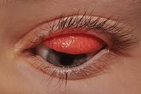
This is an inflammation of the inner lining of the upper eyelid caused by use of contact lens. The lining of the upper eye lid appears like small bubbles or stones over the surface

The condition is caused usually by trauma, and or use of soft contact lens.

Signs and symptoms includes pain, watery eyes, foreign body sensation, itching, blurry vision, et.c

Treatment involves antihistamines, steroids and lubricants.

FEMTOSECOND LASER ASSISTED CATARACT SURGERY

This is a new cataract surgery intervention that involves the use of laser beams to create micro incision on the cornea and then create a circular patch on the posterior lens capsule (a procedure called capsulectomy).

After the capsulectomy, the cataractous lens is broken down by the use of laser beams and drained out via phacoemulsification.

The procedure is done under local anesthesia and it takes about 25 to 30minutes.

RETINAL CRYOPEXY

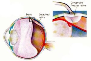
This is also called retinal cryotherapy. It is a procedure that uses freezing to seal off a retinal tear or leaking blood vessel.

This is mainly used in the treatment of retinal tear, retinal detachment and most times diabetic retinopathy.

The doctor first numbs the eye using a local anesthesia, then he places a probe that releases cold to the spot where he wants to seal off. Afterwards he releases high intensity cold to the spot for over thirty minutes.

The procedure is very safe and non invasive.

LASER PHOTOCOAGULATION

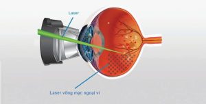
This is a Procedure that involves the use of laser beams to shrink or destroy abnormal structures in the retina..

This procedure is normally used in cases of diabetic retinopathy and other conditions that brings about neovascularization.

Although in recent times, anti VEGF drugs have been favoured in the treatment of ARMD compared to laser photocoagulation.

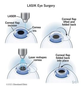
LASIK

This laser procedure involves the use of femtosecond laser or microkerotome to expose the cornea stroma and then using excimer laser to reshape the Cornea stroma inorder to improve visual acuity..

LASIK is an acronym for laser assisted in situ keratomileusis. This is a surgical procedure used to correct refractive Errors like myopia, hyperopia and astigmatism.

reference; Wikipedia

ANISOCORIA

This anisocoria is defined as an inequality in size of the pupil.

Anisocoria is usually an efferent pupillary effect.

Anisocoria can be as a result of a couple of factors which includes, trauma to the head, surgery, lesion in the visual pathway,  aneurysms, et.c.

Some signs and symptoms includes, blurry vision, photophobia, diplopia eye pain et.c

Diagnosis requires physical examination, and imaging scans .

Treatment involves treating the underlying causes

Anti – VEGF Injection

Anti – VEGF is an injection that is used to prevent or reduce the growth of abnormal blood vessels in the eye.

VEGF is vascular endothelial growth factor. This VEGF is a cytokine protein responsible for  the growth and proliferation of blood vessels. So injecting the eye with anti VEGF will  eventually reduce the proliferation of new vessels while killing off the new vessels.

This anti-VEGF is a very important technique in treating conditions like diabetic retinopathy, vascular age related macular degeneration, and other vascular eye conditions.

Some anti VEGF drugs includes pegaptanib, afligecept, ranibizumab, bevacizumab