Eagleview Eye Clinic

Archives May 2025

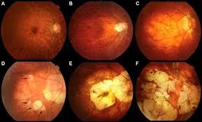
MYOPIC DEGENERATION

Myopic Degeneration

Myopic degeneration, also known as pathologic myopia, is a condition where severe nearsightedness (myopia) leads to degenerative changes in the retina. This can cause:

  • Vision loss
  • Retinal damage
  • Increased risk of retinal detachment, macular degeneration, and glaucoma

Symptoms

  1. Blurred vision: Distant objects appear blurry.
  2. Vision loss: Gradual loss of central or peripheral vision.
  3. Distorted vision: Straight lines may appear wavy or distorted.

Complications

  1. Retinal detachment: Increased risk of retinal detachment.
  2. Macular degeneration: Damage to the macula, affecting central vision.
  3. Glaucoma: Increased risk of glaucoma due to structural changes.

Management

  1. Regular eye exams: Monitoring for progression and complications.
  2. Corrective lenses: Glasses or contact lenses to correct vision.
  3. Treatment of complications: Laser therapy, surgery, or injections may be necessary to address related issues.

If you have high myopia, regular eye exams are essential to monitor for potential complications and preserve vision.
LATTICE DEGENERATION

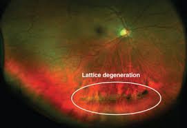
Lattice Degeneration

Lattice degeneration is a condition affecting the retina, characterized by:

  • Thinning and weakening of the retinal tissue
  • Formation of lattice-like patterns of retinal degeneration

Risks

  1. Retinal tears: Increased risk of retinal tears or holes.
  2. Retinal detachment: Potential for retinal detachment, which can lead to vision loss.

Symptoms

  1. Often asymptomatic: Lattice degeneration may not cause noticeable symptoms.
  2. Floaters or flashes: Some people may experience floaters or flashes of light.

Management

  1. Regular eye exams: Monitoring for progression or complications.
  2. Laser photocoagulation: Treatment to strengthen the retina and prevent retinal detachment.
  3. Retinal detachment repair: Surgery may be necessary if retinal detachment occurs.

If you have lattice degeneration, regular eye exams with a retina specialist are crucial to monitor the condition and prevent potential complications.

POSTERIOR STAPHYLOMA

Posterior Staphyloma

Posterior staphyloma is a condition where the sclera (the white part of the eye) becomes thin and bulges outward, typically in the posterior segment of the eye. This can be associated with:

  • High myopia (nearsightedness)
  • Retinal degeneration
  • Increased risk of retinal detachment

Symptoms

  1. Vision problems: Blurred vision, distorted vision, or blind spots.
  2. Retinal complications: Retinal detachment, retinal tears, or macular degeneration.

Causes

  1. High myopia: Severe nearsightedness can lead to scleral thinning.
  2. Genetic predisposition: Family history may play a role.

Management

  1. Regular eye exams: Monitoring for complications.
  2. Treatment of associated conditions: Managing high myopia, retinal detachment, or other related issues.
  3. Surgical interventions: May be necessary to address retinal detachment or other complications.

If you have concerns about posterior staphyloma or high myopia, consult an eye care professional for personalized advice and management.

FLOATERS

Floaters are small, dark spots that appear to float in front of your eyes. They’re caused by tiny pieces of the eye’s gel-like substance (vitreous) that break down and clump together.

Symptoms

  1. Spots or threads: Seeing small, dark spots or thread-like structures.
  2. Movement: Floaters move as your eyes move.
  3. More noticeable: Floaters are more noticeable when looking at a bright, uniform background.

Causes

  1. Aging: Floaters are a common age-related phenomenon.
  2. Vitreous detachment: As we age, the vitreous gel can shrink and detach from the retina.
  3. Eye inflammation: Certain conditions can cause inflammation that leads to floaters.

When to Seek Medical Attention
If you experience:

  • Sudden increase in floaters: A sudden shower of floaters.
  • Flashes of light: Seeing flashes of light.
  • Vision loss: Any loss of vision.

Consult an eye care professional if you’re concerned about floaters or if you experience any unusual symptoms.

Ocular Ischemic Syndrome.

Ocular ischemic syndrome is a rare condition caused by severe carotid artery stenosis or occlusion, leading to reduced blood flow to the eye. This can result in:

  • Gradual vision loss
  • Pain in the eye
  • Retinal damage

Causes

  1. Carotid artery disease: Narrowing or blockage of the carotid artery.
  2. Atherosclerosis: Buildup of plaque in the arteries.

Symptoms

  1. Vision loss: Gradual or sudden loss of vision.
  2. Eye pain: Pain or discomfort in the affected eye.
  3. Retinal hemorrhages: Bleeding in the retina.

Treatment

  1. Carotid endarterectomy: Surgery to remove plaque from the carotid artery.
  2. Angioplasty and stenting: Procedures to improve blood flow.
  3. Management of underlying conditions: Controlling hypertension, diabetes, and other risk factors.

If you suspect ocular ischemic syndrome, seek immediate medical attention from an eye care professional or a vascular specialist. Prompt treatment can help prevent further vision loss.

GIANT CELL ARTERITIS

Giant cell arteritis is an inflammation of blood vessels, typically affecting the head, especially the temples. Symptoms include:
– Headaches
– Scalp tenderness
– Jaw pain
– Vision problems
– Fatigue

If left untreated, it can lead to serious complications like vision loss. Treatment usually involves corticosteroids. Consult a healthcare professional for proper diagnosis and care.

MEGALOCORNEA


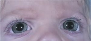
Megalocornea is a rare, non-progressive condition characterized by an abnormally large cornea, typically greater than 13 mm in diameter. This condition is often present at birth and can occur in one or both eyes.

Characteristics


1. _Large corneal diameter_: The cornea is significantly larger than average.


2. _Normal corneal thickness_: Despite its size, the cornea is usually of normal thickness.


3. _Often associated with other ocular features_: Megalocornea can be associated with other eye abnormalities.

Potential Implications


1. _Refractive errors_: Patients may experience nearsightedness, farsightedness, or astigmatism.


2. _Increased risk of glaucoma_: Regular eye exams are essential to monitor intraocular pressure.

*LManagement


1. _Regular eye exams_: Monitoring for potential complications.


2. _Corrective lenses_: Glasses or contact lenses to correct refractive errors.


3. _Surgical interventions_: May be necessary to address associated conditions.

If you have concerns about megalocornea or its implications, consult an eye care professional for personalized advice and management.

HYPEROPIA


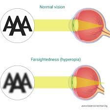
Hyperopia, also known as farsightedness, is a common vision condition where distant objects are seen clearly, but nearby objects appear blurry. This occurs when the eyeball is too short or the cornea is too flat, causing light to focus behind the retina instead of directly on it.

Symptoms


1. _Blurry near vision_: Difficulty seeing objects up close.


2. _Eye strain_: Discomfort or fatigue after reading or other near-work activities.


3. _Headaches_: Frequent headaches due to prolonged eye strain.

Correction


1. _Glasses_: Convex lenses to converge light rays.


2. _Contact lenses_: Convex lenses to correct vision.


3. _Refractive surgery_: Procedures like LASIK to reshape the cornea.

If you’re experiencing symptoms, consult an eye care professional for a comprehensive eye exam and personalized guidance.

ORBITAL CELLULITIS

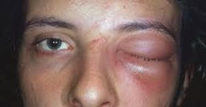
Orbital cellulitis is a serious infection of the tissues surrounding the eye, posterior to the orbital septum. It’s often caused by bacterial infections, typically Staphylococcus aureus or Streptococcus pneumoniae.

Symptoms

  1. Severe swelling: Redness, swelling, and warmth around the eye.
  2. Pain: Significant pain, especially with eye movement.
  3. Vision problems: Blurred vision, double vision, or loss of vision.
  4. Fever: High fever, often accompanied by chills.
  5. Proptosis: Bulging of the eye.

Causes

  1. Sinusitis: Spread of infection from sinuses.
  2. Trauma: Direct inoculation of bacteria through a wound.
  3. Hematogenous spread: Infection spread through the bloodstream.

Complications

  1. Vision loss: Permanent vision loss if left untreated.
  2. Intracranial complications: Infection can spread to the brain, leading to meningitis or abscesses.
  3. Cavernous sinus thrombosis: Blood clot in the cavernous sinus.

Treatment

  1. Antibiotics: Intravenous antibiotics to treat the bacterial infection.
  2. Hospitalization: Often requires hospitalization for close monitoring.
  3. Surgery: May be necessary to drain abscesses or relieve pressure.

If you suspect orbital cellulitis, seek immediate medical attention from a healthcare professional or an optometrist. Prompt treatment is crucial to prevent serious complications.

PRESEPTAL CELLULITIS


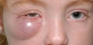
Preseptal cellulitis is an infection of the eyelid and surrounding skin, anterior to the orbital septum. It’s often caused by bacterial infections, typically Staphylococcus aureus or Streptococcus pneumoniae.

Symptoms


1. _Swelling_: Redness and swelling of the eyelid.


2. _Pain_: Tenderness to the touch.


3. _Warmth_: Affected area may feel warm.


4. _Fever_: May be present in some cases.


Causes


1. _Insect bites_: Bug bites can introduce bacteria.


2. _Skin trauma_: Cuts, scratches, or other injuries.


3. _Sinusitis_: Spread of infection from sinuses.

Treatment


1. _Antibiotics_: Oral or intravenous antibiotics to treat the bacterial infection.


2. _Warm compresses_: May help reduce swelling.

Complications


1. _Orbital cellulitis_: Infection can spread posteriorly, leading to more severe orbital cellulitis.


2. _Vision problems_: Untreated preseptal cellulitis can potentially lead to complications affecting vision.

If you’re experiencing symptoms or concerns, consult a healthcare professional for proper evaluation and treatment.