Eagleview Eye Clinic

Archives May 2025

MICROCORNEA


Microcornea is a rare congenital condition where the cornea is abnormally small, typically less than 10 mm in diameter. This can occur in one or both eyes.

Characteristics


1. _Small corneal diameter_: The cornea is smaller than usual.


2. _Associated conditions_: Microcornea can be associated with other ocular or systemic abnormalities.

Potential Complications


1. _Hyperopia_: Microcornea can lead to farsightedness due to the shorter axial length.


2. _Glaucoma_: Increased risk of developing glaucoma.


3. _Other eye abnormalities_: Microcornea may be associated with other ocular malformations.

Diagnosis


1. _Ophthalmological examination_: Measurement of corneal diameter.


2. _Imaging tests_: Ultrasound or other imaging modalities to assess the eye.

Management


1. _Corrective lenses_: Glasses or contact lenses to correct refractive errors.


2. _Monitoring_: Regular eye exams to monitor for potential complications.


3. _Surgical interventions_: In some cases, surgery may be necessary to address associated conditions.

If you’re concerned about microcornea or have questions, consult an eye care professional for personalized guidance.

PTOSIS

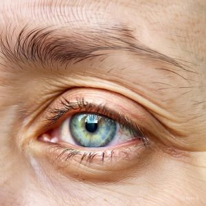
Ptosis

Ptosis is a medical condition characterized by drooping or falling of the upper eyelid. This can affect one or both eyes and may be present at birth (congenital) or develop later in life (acquired).

Types

  1. Congenital ptosis: Present at birth, often due to underdevelopment of the levator muscle.
  2. Acquired ptosis: Develops later in life, potentially caused by:
    1. Aging
    2. Injury or trauma
    3. Neurological conditions (e.g., myasthenia gravis, stroke)
    4. Muscle diseases

Symptoms

  1. Drooping eyelid: Visible sagging of the upper eyelid.
  2. Vision obstruction: In severe cases, the drooping lid may obstruct vision.
  3. Compensatory mechanisms: Individuals might tilt their head back or raise their eyebrows to see.

Treatment

  1. Surgery: Often recommended to repair or strengthen the levator muscle.
  2. Non-surgical options: In some cases, ptosis crutches or Botox injections might be used.

If you’re experiencing symptoms or concerns, consult an eye care professional or a healthcare provider for proper evaluation and guidance.

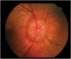
PAPILLOEDEMA


Papilloedema is a medical condition characterized by swelling of the optic disc, which is the point on the retina where the optic nerve fibers exit the eye. This swelling is usually caused by increased intracranial pressure (ICP).

Causes


1. _Increased intracranial pressure_: Conditions like brain tumors, meningitis, or idiopathic intracranial hypertension can cause papilloedema.


2. _Head trauma_: Severe head injuries can lead to increased ICP and papilloedema.

Symptoms


1. _Visual disturbances_: Transient vision loss, double vision, or blind spots.


2. _Headaches_: Often worse in the morning.


3. _Nausea and vomiting_: May occur due to increased ICP.

Complications


1. _Vision loss_: Untreated papilloedema can lead to permanent vision loss.


2. _Optic nerve damage_: Prolonged swelling can damage the optic nerve.

Diagnosis


1. _Ophthalmoscopy_: Examination of the optic disc to detect swelling.


2. _Imaging tests_: MRI or CT scans to identify underlying causes.

Treatment


1. _Addressing underlying cause_: Treating the underlying condition causing increased ICP.


2. _Monitoring_: Regular eye exams to track optic disc swelling and vision.

If you’re experiencing symptoms or concerns, consult an eye care professional or a healthcare provider for proper evaluation and guidance.

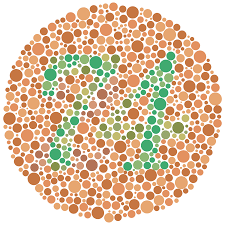
COLOUR VISION DEFICIENCY


Color vision deficiency, also known as color blindness, is a condition where a person has difficulty perceiving certain colors. This is usually due to a genetic mutation affecting the light-sensitive cells (photoreceptors) in the retina.

Types


1. _Red-green color blindness_: Most common type, where individuals struggle to distinguish between red and green colors.


2. _Monochromacy_: Rare condition where a person sees the world in shades of gray only.


3. _Tritanopia_: Difficulty perceiving blue colors.

Causes


1. _Genetics_: Inherited trait, more common in males.


2. _Age-related_: Can occur due to aging.


3. _Eye diseases_: Certain conditions like cataracts or age-related macular degeneration.

Effects


1. _Difficulty with color distinction_: Challenges in everyday tasks, like selecting ripe fruit or understanding color-coded information.


2. _Adaptation_: Many people develop coping strategies.

Diagnosis


1. _Ishihara test_: A common test using dot patterns to identify color vision deficiency.


2. _Other tests_: Like the Hardy-Rand-Rittler test or the City University Color Vision Test.

If you’re concerned about color vision deficiency, consult an eye care professional for proper evaluation and guidance.

CHARLES BONNET SYNDROME

Charles Bonnet Syndrome

Charles Bonnet Syndrome (CBS) is a condition where people with significant vision loss experience vivid visual hallucinations. These hallucinations can range from simple patterns to complex images, such as people, animals, or objects.

Characteristics

  1. Visual hallucinations: CBS hallucinations are typically visual and can be very vivid.
  2. Vision loss: CBS typically occurs in people with significant vision loss, often due to age-related macular degeneration, cataracts, or other eye conditions.
  3. Insight: People with CBS often have insight into the fact that their hallucinations are not real.

Symptoms

  1. Hallucinations: Can be simple (e.g., patterns, shapes) or complex (e.g., people, animals).
  2. Variable frequency: Hallucinations can occur frequently or infrequently.
  3. Variable duration: Hallucinations can last from seconds to hours.

Management

  1. Reassurance: Educating patients that CBS is a recognized condition can be helpful.
  2. Coping strategies: Techniques like closing eyes or looking away can help manage hallucinations.
  3. Addressing underlying conditions: Managing underlying eye conditions can help alleviate symptoms.

If you’re experiencing symptoms or concerns, consult an eye doctor or a healthcare professional for proper diagnosis and guidance.

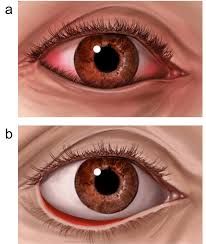
ECTROPION


Ectropion is a condition where the eyelid turns outward, often causing the eyelid to sag away from the eye. This can lead to:

1. _Eye dryness_: Exposure of the eye surface.


2. _Irritation_: Redness, tearing, and discomfort.


3. _Increased risk of infection_: Exposure to the environment.

Causes include:

1. _Aging_: Weakening of eyelid muscles.


2. _Facial paralysis_: Conditions like Bell’s palsy.


3. _Scarring_: From injury or surgery.

Treatment may involve surgery to tighten or reposition the eyelid.

If you’re experiencing symptoms, consult an eye doctor for proper diagnosis and treatment.

ENTROPION

Entropion

Entropion is a condition where the eyelid rolls inward, causing the eyelashes to rub against the cornea. This can lead to:

  1. Eye irritation: Discomfort, redness, and tearing.
  2. Corneal damage: Potential scarring or ulcers.

Causes include:

  1. Aging: Weakening of eyelid muscles.
  2. Scarring: From injury or infection.
  3. Muscle weakness: Certain neurological conditions.

Treatment often involves surgery to tighten or reposition the eyelid. If you’re experiencing symptoms, consult an eye doctor for proper diagnosis and treatment.

ETOPIA LENTIS

Ectopia Lentis

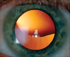
Ectopia lentis is a rare condition where the lens of the eye is displaced or malpositioned. This can occur due to various reasons, including:

Causes

  1. Trauma: Blunt or penetrating eye injury can cause the lens to become dislocated.
  2. Genetic conditions: Certain genetic disorders, such as Marfan syndrome, Ehlers-Danlos syndrome, or homocystinuria, can increase the risk of ectopia lentis.
  3. Weakening of the zonular fibers: The zonular fibers hold the lens in place. Weakening or damage to these fibers can cause the lens to become dislocated.

Symptoms

  1. Blurred vision: Displacement of the lens can cause vision problems.
  2. Double vision: Ectopia lentis can cause double vision or ghosting.
  3. Eye pain: In some cases, ectopia lentis can cause eye pain or discomfort.

Treatment

  1. Surgery: Surgical repositioning or removal of the lens may be necessary.
  2. Corrective lenses: Glasses or contact lenses may be prescribed to correct vision problems.

If you suspect you have ectopia lentis or are experiencing symptoms, consult an eye doctor for proper diagnosis and treatment.

CORNEA DYSTROPHIES

Corneal Dystrophy

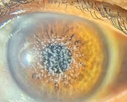
Corneal dystrophy is a group of genetic eye disorders that affect the cornea, leading to progressive vision loss. It’s characterized by:

  1. Abnormal corneal changes: Clouding, thinning, or scarring of the cornea.
  2. Gradual vision loss: Vision becomes increasingly blurred or distorted.
  3. Inherited: Often passed down through families.

Types include Fuchs’ dystrophy, keratoconus, and others.

Treatment may involve medication, surgery, or corneal transplant.

Regular eye exams are essential for monitoring and managing the condition.

CORNEA SCAR

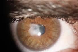
A corneal scar is an opacity or mark on the cornea, often caused by injury, infection, or surgery, affecting vision and potentially causing blurred or distorted vision.
Treatment options for corneal scars depend on the severity and location of the scar. They may include:


Treatment options for corneal scars depend on the severity and location of the scar. They may include:

1. Observation: Monitoring the scar for changes.


2. Medications: Using topical medications to reduce inflammation.


3. Corneal scraping or smoothing: Removing or smoothing the scar tissue.


4. Phototherapeutic keratectomy (PTK): Using laser treatment to remove scar tissue.


5. Corneal transplant: Replacing the damaged cornea with a donor cornea.