Eagleview Eye Clinic

Archives July 2025

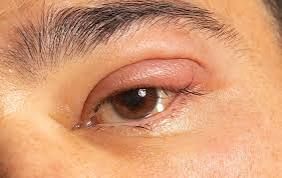
NECROTIZING SCLERITIS

Necrotizing Scleritis

Necrotizing scleritis is a:

  • Severe inflammatory eye condition
  • Characterized by:
    1. Scleral inflammation: Inflammation of the white part of the eye
    2. Tissue necrosis: Death of scleral tissue
    3. Vision-threatening: Can lead to vision loss if untreated

Symptoms

  1. Severe eye pain: Often described as deep, boring pain
  2. Redness: Inflammation of the sclera
  3. Vision loss: Blurred vision or decreased vision

Treatment

  1. Immunosuppressive medications: To control inflammation
  2. Corticosteroids: May be used to reduce inflammation
  3. Surgical intervention: In some cases

If you suspect necrotizing scleritis, consult an eye care professional or rheumatologist for prompt diagnosis and treatment to prevent vision loss.

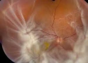
PROLIFERATIVE VITREORETINOPATHY

Proliferative Vitreoretinopathy (PVR)

Proliferative vitreoretinopathy (PVR) is a:

  • Complication of retinal detachment or eye injury
  • Characterized by:
    1. Scarring: Formation of fibrotic membranes in the vitreous and retina
    2. Retinal traction: Contraction of scar tissue, leading to retinal detachment or vision loss

Causes

  1. Retinal detachment: Previous detachment or surgery
  2. Eye injury: Trauma or inflammation

Treatment

  1. Surgery: Vitrectomy and membrane peeling to remove scar tissue
  2. Complex retinal surgery: May require multiple procedures

If you have PVR, consult a retina specialist for personalized advice and treatment to manage the condition and preserve vision.

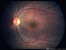
FUNDUS ALBIPUNCTATUS

Fundus Albipunctatus

Fundus albipunctatus is a:

  • Rare, inherited condition affecting the retina
  • Characterized by:
    1. White or yellowish dots: In the retina
    2. Night blindness: Difficulty seeing in low light
    3. Variable visual impairment: May experience reduced vision

Causes

  1. Genetic mutation: Affecting the RDH5 gene

Treatment

  1. No cure: Currently, no cure available
  2. Supportive care: Regular eye exams, low vision aids

If you have fundus albipunctatus, consult an eye care professional or retina specialist for personalized advice and support to manage vision loss.

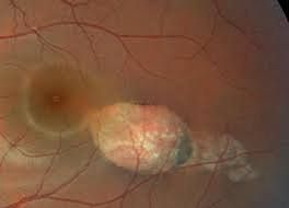
TORPEDO MACULOPATHY

Torpedo Maculopathy

Torpedo maculopathy is a:

  • Rare, congenital condition affecting the macula
  • Characterized by a distinctive “torpedo-shaped” lesion

Symptoms

  1. Vision loss: Variable, often mild to moderate
  2. Visual field defects: Blind spots or distorted vision

Characteristics

  1. Lesion shape: Torpedo-shaped or oval-shaped lesion in the macula
  2. Retinal pigment epithelium (RPE) changes: Abnormalities in the RPE layer

Treatment

  1. Monitoring: Regular eye exams to track progression
  2. No treatment: Often not necessary, but low vision aids may be helpful

If you have torpedo maculopathy, consult an eye care professional or retina specialist for personalized advice and monitoring to preserve vision.

STAGARDT DISEASE

Stargardt Disease

Stargardt disease is a:

  • Genetic eye disorder affecting the macula
  • Leading cause of inherited juvenile macular degeneration

Symptoms

  1. Vision loss: Central vision loss, blurred vision
  2. Color vision: Difficulty distinguishing colors
  3. Night vision: Impaired adaptation to darkness

Characteristics

  1. Macular degeneration: Damage to the macula, causing vision loss
  2. Lipofuscin accumulation: Abnormal buildup of lipofuscin in the retina

Treatment

  1. No cure: Currently, no cure available
  2. Supportive care: Regular eye exams, low vision aids
  3. Research: Ongoing studies on potential treatments, such as gene therapy

If you have Stargardt disease, consult an eye care professional or retina specialist for personalized advice and support to manage vision loss.

INFECTIOUS RETINITIS

Infectious Retinitis

Infectious retinitis is inflammation of the retina caused by:

  • Viral, bacterial, fungal, or parasitic infections

Types

  1. Cytomegalovirus (CMV) retinitis: Common in immunocompromised individuals
  2. Toxoplasmosis retinochoroiditis: Caused by Toxoplasma gondii parasite
  3. Herpes simplex or varicella-zoster virus retinitis: Viral infections

Symptoms

  1. Vision loss: Blurred vision, blind spots
  2. Floaters: Dark spots or floaters in vision
  3. Eye pain: Redness, discomfort

Treatment

  1. Antiviral medications: For viral infections
  2. Antiparasitic medications: For toxoplasmosis
  3. Antibiotics: For bacterial infections
  4. Intravitreal injections: Direct medication delivery into the eye

If you suspect infectious retinitis, consult an eye care professional or retina specialist for proper diagnosis and treatment to preserve vision.

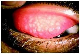
TRACHOMA

Trachoma

Trachoma is a:

  • Bacterial infection (Chlamydia trachomatis) affecting the eyes
  • Leading cause of preventable blindness worldwide

Symptoms

  1. Eye irritation: Redness, itching, discharge
  2. Follicles: On the underside of the upper eyelid
  3. Scarring: Can lead to vision loss and blindness

Transmission

  1. Direct contact: With infected eye discharge
  2. Indirect contact: Through contaminated objects or flies

Prevention

  1. Hygiene: Proper handwashing and face washing
  2. Sanitation: Improved access to clean water and sanitation
  3. Antibiotics: Mass treatment in endemic areas

Treatment

  1. Antibiotics: Azithromycin or tetracycline
  2. Surgery: To correct eyelid deformities or trichiasis (eyelashes rubbing against the cornea)

If you suspect trachoma, consult an eye care professional for proper diagnosis and treatment. Early intervention can prevent long-term vision damage.

OPTIC NERVE GLIOMA AND MENINGIOMA

Optic Nerve Glioma and Meningioma

Optic Nerve Glioma

  1. Type of tumor: Benign or low-grade tumor arising from glial cells
  2. Association: Often linked to neurofibromatosis type 1 (NF1)
  3. Symptoms: Vision loss, proptosis (bulging eye), or optic nerve damage

Meningioma

  1. Type of tumor: Typically benign tumor arising from meninges (protective membranes around brain and spinal cord)
  2. Location: Can occur near optic nerve or other areas of the brain
  3. Symptoms: Vision loss, double vision, or other neurological symptoms

Treatment

  1. Monitoring: Regular imaging and eye exams
  2. Surgery: To remove tumor or relieve pressure
  3. Radiation therapy: In some cases

If you suspect an optic nerve glioma or meningioma, consult a neuro-ophthalmologist or neurosurgeon for proper diagnosis and treatment. Early detection is crucial for effective management.

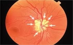
OPTIC DISC DRUSEN

Optic Disc Drusen

Optic disc drusen are:

  • Small, calcified deposits that accumulate in the optic disc
  • Can be congenital or develop over time
  • May cause visual field defects or optic nerve damage

Symptoms

  1. Visual field loss: Gradual loss of peripheral or central vision
  2. No symptoms: Often asymptomatic, discovered during routine eye exams

Diagnosis

  1. Fundus examination: Ophthalmoscopy or fundus photography
  2. Imaging tests: Ultrasound, CT, or OCT (optical coherence tomography)

Treatment

  1. Monitoring: Regular eye exams to track progression
  2. No treatment: Often not necessary, but addressing related conditions may be required

If you have optic disc drusen, consult an eye care professional for personalized advice and monitoring to preserve vision and eye health.

DACRYOADENITIS

Dacryoadenitis

Dacryoadenitis is inflammation of the lacrimal gland, which can be:

  • Acute or chronic
  • Caused by infection, autoimmune disorders, or blockage

Symptoms

  1. Swelling: Upper eyelid swelling, often on the outer side
  2. Pain: Tenderness or pain in the area of the lacrimal gland
  3. Redness: Inflammation of the eyelid
  4. Excessive tearing: Or decreased tearing in chronic cases

Causes

  1. Infections: Bacterial or viral
  2. Autoimmune disorders: Such as sarcoidosis or Sjögren’s syndrome
  3. Blockage: Of the lacrimal gland ducts

Treatment

  1. Warm compresses: To relieve symptoms
  2. Antibiotics: If bacterial infection is present
  3. Anti-inflammatory medications: To reduce swelling and pain
  4. Further testing: May be needed to determine underlying cause

If you have dacryoadenitis, consult an eye care professional for proper diagnosis and treatment to alleviate symptoms and address the underlying cause.