Full-field ERG tests the entire retina’s electrical response to light. You get electrodes on the cornea or skin, dark-adapt for ~20 mins, then flashes of light stimulate rods and cones. It records a-waves (photoreceptor activity) and b-waves (inner retinal response). Used to diagnose widespread retinal diseases like retinitis pigmentosa or Leber congenital amaurosis. Results show overall retinal function, not localized areas.
CORNEA DECOMPENSATION
👁️ Corneal decompensation happens when the endothelium (the inner layer of the cornea) fails to pump out fluid, causing corneal swelling (edema), cloudiness, and blurry vision.
- Causes:
- Fuchs’ endothelial dystrophy (most common)
- Trauma (surgical or accidental)
- Long-term contact lens wear
- Glaucoma or high eye pressure
- Inflammation or infection
- Symptoms:
- Hazy or blurry vision (often worse in the morning)
- Glare and light sensitivity
- Eye discomfort or pain in advanced cases
- Halos around lights
- Stages:
- Early: Mild swelling, subtle visual changes
- Moderate: Noticeable edema, thickened cornea
- Advanced: Bullae formation (epithelial blisters), pain, and risk of infection
- Treatment:
- Mild: Hypertonic saline drops/ointment to draw out fluid
- Moderate: Reducing contributing factors (e.g., pressure control in glaucoma)
- Severe: Endothelial keratoplasty (DSEK, DSAEK, or DMEK) — partial corneal transplant replacing only the damaged endothelial layer
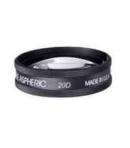
20D LENS
👓 Here’s more on a 20D lens:
- Magnification: A +20D lens provides approximately 20x magnification.
- Field of view: Offers a relatively wide field, useful for examining the retina.
- Use: Commonly used in ophthalmology for:
- Indirect ophthalmoscopy (with a slit lamp or head-mounted scope).
- Examining the posterior segment (retina, macula, etc.).
- Type: Typically a handheld, convex lens made of glass or acrylic.
- Working distance: Around 5 cm (2 inches) from the patient’s eye.
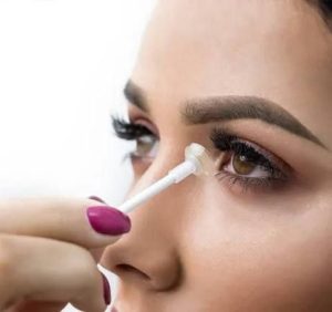
CONTACT LENS APPLICATOR
A contact lens applicator 😎 is a handy tool that helps you put in (and sometimes take out) contact lenses, especially if you’re a newbie or have dexterity issues. Types include:
- Sucker/plucker: A small tool with a suction cup that grips the lens.
- Finger-tip applicator: A guide that fits on your finger to help position the lens.
They’re super useful if:
- You’re struggling to handle tiny lenses
- You have arthritis or finger issues
- You’re helping someone else with lenses (like kids)
DIAMOX
👨⚕️ Diamox (Acetazolamide) is a carbonic anhydrase inhibitor.
Uses:
- Glaucoma: Reduces intraocular pressure
- Edema: Treats fluid retention
- Altitude sickness: Prevents and treats symptoms
- Epilepsy: Adjunct therapy
Eye-related uses:
- Iocular pressure reduction: Glaucoma treatment
- Cystoid macular edema: Treats fluid buildup
Side effects:
- Numbness/tingling: Hands, feet, or face
- Dizziness: May affect daily activities
- Increased urination: Monitor hydration
LISAMINE GREEN STAINING
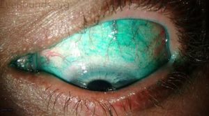
👨⚕️ Lissamine Green staining test assesses ocular surface damage, particularly conjunctival epithelial damage.
Procedure:
- Apply anesthetic drops (optional)
- Place Lissamine Green drops in eye
- Examine under white or blue light
Interpretation:
- Staining pattern: Indicates damage location/severity
- Scoring systems: Grade staining (e.g., Oxford scale)
Uses:
- Dry eye disease: Evaluates conjunctival staining
- Ocular surface disorders: Identifies damaged areas
- Conjunctival disorders: Assesses inflammation
FLOURESCEIN ANGIOGRAPHY
👨⚕️ Fluorescein angiography is a diagnostic test that uses fluorescein dye and a specialized camera to visualize the retinal blood vessels.
Key uses:
- Diabetic retinopathy: Detects leakage, neovascularization
- Macular degeneration: Identifies abnormal blood vessels
- Retinal vascular occlusions: Diagnoses blockages
- Tumors: Highlights abnormal vessels
Procedure:
- Inject fluorescein dye into arm vein
- Take photographs as dye circulates
- Capture early and late-phase images
Findings:
- Leakage: Dye escape from vessels
- Blockage: Dark areas (hypofluorescence)
- Neovascularization: Abnormal vessels
Precautions:
- Nausea, yellow skin discoloration (temporary)
- Allergic reactions (rare)
- Pregnancy/breastfeeding: Use cautiously
REFRACTION
🔍 Refraction is the bending of light as it passes through different mediums (e.g., air, lens, cornea). In the eye, it focuses light onto the retina.
Types:
- Emmetropia: Normal vision.
- Myopia (nearsightedness): Blurred distance vision.
- Hyperopia (farsightedness): Blurred near vision.
- Astigmatism: Distorted vision.
- Presbyopia: Age-related near vision loss.
Tests:
- Visual acuity: Snellen chart.
- Autorefraction: Objective measurement.
- Subjective tests: Lens power adjustment.
Corrections:
- Glasses/contact lenses: Adjust refractive power.
- Surgery: LASIK, PRK, etc.
MYDRIATIC DRUGS
👁️ Mydriatic drugs dilate the pupil (mydriasis) by relaxing the iris sphincter muscle. Used for exams, surgery, or treating inflammation.
Common examples:
- Phenylephrine (α-1 agonist)
- Effect: Fast (30–60 min), short (3–6h).
- Use: Diagnostic exams.
- Risk: Hypertension, arrhythmia.
- Tropicamide (anticholinergic)
- Effect: 30–45 min, lasts 4–6h.
- Use: Routine dilation.
- Risk: Blurred vision, photophobia.
- Atropine (strong, long-acting)
- Effect: 1–2 weeks.
- Use: Uveitis, amblyopia.
- Risk: Prolonged blur, dry mouth.
- Cyclopentolate
- Effect: 24–48h.
- Use: Pediatric exams.
- Risk: Behavioral changes in kids.
Precautions:
- Avoid driving until pupils return to normal.
- Glaucoma risk: Check IOP.
- Monitor elderly/kids.
- Side effects: Headache, stinging, dry eyes.