Eagleview Eye Clinic

Tag eye care professionals

CORNEA

The cornea is the transparent, avascular (without blood vessels), dome-shaped surface at the front of the eye. It plays a crucial role in refracting (bending) light and protecting the eye.

Key characteristics:

  1. Transparency: The cornea is clear and transparent, allowing light to pass through.
  2. Structure: It consists of five layers:
    • Epithelium (outermost layer)
    • Bowman’s layer
    • Stroma (thickest layer)
    • Descemet’s membrane
    • Endothelium (innermost layer)
  3. Function: The cornea:
    • Refracts light, accounting for most of the eye’s focusing power
    • Protects the eye from external damage
    • Maintains the shape of the eye
  4. Sensitivity: The cornea is highly sensitive to touch, temperature, and chemicals.
  5. Nourishment: The cornea receives nutrients and oxygen from tears and the aqueous humor.

Importance: The cornea is essential for clear vision, and any damage, disease, or irregularity can significantly impact visual acuity.

SCLERA

The sclera, also known as the white of the eye, is the opaque, fibrous, and protective outer layer of the eyeball. It provides structure, protection, and shape to the eye.

Key characteristics:

  1. Composition: The sclera is made of dense, collagenous connective tissue.
  2. Location: It covers the posterior five-sixths of the eyeball, with the cornea covering the anterior one-sixth.
  3. Function: The sclera:
    • Provides protection to the intraocular contents
    • Maintains the shape of the eyeball
    • Serves as an attachment site for the extraocular muscles that control eye movement
  4. Appearance: The sclera is typically white or off-white in color, but it can appear differently in certain conditions.

Importance: The sclera plays a vital role in maintaining the integrity and function of the eye, and its health is essential for preserving vision.

BLEB

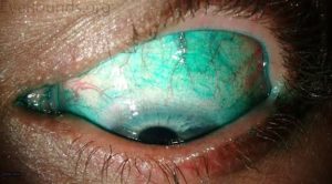
A bleb is a blister-like structure that forms on the surface of the eye, typically as a result of a surgical procedure, such as trabeculectomy. In the context of glaucoma surgery, a bleb is a fluid-filled reservoir that forms under the conjunctiva, allowing excess fluid to drain from the eye and reducing intraocular pressure.

Characteristics of a bleb:

  • Fluid-filled
  • Located under the conjunctiva
  • Can be diffuse or localized
  • May be elevated or flat

The presence and characteristics of a bleb can indicate the success of the surgery and the effectiveness of IOP reduction.

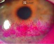
NEUROPARALYTIC KERATITIS

Neuroparalytic keratitis is a condition where the cornea becomes inflamed or damaged due to nerve dysfunction or paralysis, often resulting from:

  • Facial nerve palsy (e.g., Bell’s palsy)
  • Trigeminal nerve damage
  • Trauma
  • Stroke
  • Neurological disorders (e.g., multiple sclerosis)

This condition can lead to:

  • Decreased corneal sensation
  • Reduced blinking
  • Exposure keratopathy
  • Increased risk of corneal ulcers

Symptoms may include:

  • Eye pain
  • Redness
  • Blurred vision
  • Dryness

Treatment aims to:

  • Protect the cornea
  • Promote healing
  • Manage symptoms

Options may include:

  • Lubricating eye drops
  • Protective eyewear
  • Tarsorrhaphy (surgical eyelid closure)
  • Other supportive measures.
ORBITAL TUMOUR REMOVAL

Orbital tumor removal is a surgical procedure to remove abnormal growths or tumors within the orbit (the bony cavity surrounding the eye). The goal is to:

  • Eliminate the tumor
  • Preserve vision and eye function
  • Improve appearance
  • Relieve symptoms (e.g., pain, double vision)

Types of orbital tumors:

  • Benign (non-cancerous): e.g., hemangiomas, neurofibromas
  • Malignant (cancerous): e.g., lymphoma, rhabdomyosarcoma

Surgical approaches:

  • Anterior orbitotomy: For tumors located in the front part of the orbit.
  • Lateral orbitotomy: For tumors located on the side of the orbit.
  • Cranio-orbital approach: For tumors extending into the cranial cavity.

Techniques:

  • Open surgery: Traditional approach with incisions.
  • Endoscopic surgery: Minimally invasive approach using small incisions and a camera.

Post-operative care:

  • Monitoring for complications (e.g., bleeding, infection)
  • Managing pain and swelling
  • Follow-up appointments for recovery assessment

The specific approach and technique depend on the tumor’s location, size, and type, as well as the patient’s overall health.

LEVATOR RESECTION

Levator resection is a surgical technique used to correct ptosis (drooping eyelid) by strengthening the levator palpebrae superioris muscle, which is responsible for eyelid elevation. Here’s a more detailed overview:

Indications:

  • Congenital or acquired ptosis
  • Mild to moderate ptosis
  • Good levator function

Procedure:

  1. The surgeon makes an incision in the eyelid crease.
  2. The levator muscle and tendon are identified and resected (a portion is removed).
  3. The resected muscle is then reattached to the eyelid, effectively shortening it and improving eyelid elevation.

Goals:

  • Improve eyelid position and symmetry
  • Enhance visual field
  • Restore normal appearance

Types:

  • Müllerectomy: A variation involving resection of Müller’s muscle, which also contributes to eyelid elevation.

Post-operative care:

  • Eye drops and ointments may be prescribed to aid healing and reduce inflammation.
  • Patients are usually advised to avoid heavy lifting, bending, or strenuous activities.

Potential risks and complications:

  • Undercorrection or overcorrection
  • Bleeding or hematoma
  • Infection
  • Lagophthalmos (incomplete eyelid closure)

Levator resection is a common and effective procedure for correcting ptosis, but the outcome depends on individual factors, such as the severity of ptosis and the surgeon’s expertise.

TARSORRHAPHY

Tarsorrhaphy is a surgical procedure that involves stitching or suturing the eyelids together, either partially or completely, to protect the eye and promote healing. This procedure can be temporary or permanent and is often used to address conditions such as:

  1. Exposure keratopathy (dryness or damage to the cornea due to incomplete eyelid closure)
  2. Neuroparalytic keratitis (inflammation of the cornea due to nerve damage)
  3. Facial paralysis or weakness

By limiting eyelid movement, tarsorrhaphy helps shield the eye from external factors, reduces discomfort, and supports the healing process.

FRONTALIS SLING

A frontalis sling is a surgical procedure used to:

  1. Treat ptosis: Correct droopy eyelids by suspending the eyelid from the frontalis muscle.
  2. Improve eyelid elevation: Enhance eyelid movement and symmetry.

The procedure involves:

  1. Sling material: Using a material (e.g., silicone, fascia lata) to connect the eyelid to the frontalis muscle.
  2. Customized approach: Tailored to individual needs and anatomy.

Frontalis sling surgery can significantly improve eyelid function and appearance.

MULLERECTOMY

Mullerectomy is a surgical procedure that involves the removal or resection of a portion of the Müller’s muscle in the eyelid. It’s often performed to:

  1. Treat ptosis: Correct droopy eyelids by strengthening the eyelid’s lifting mechanism.
  2. Improve eyelid function: Enhance eyelid elevation and symmetry.

The procedure is typically done by an oculoplastic surgeon and can be tailored to individual needs.

TEAR DUCT SURGERY

Tear duct surgery, also known as dacryocystorhinostomy (DCR), is a procedure to:

  1. Restore tear drainage: Creating a new pathway for tears to flow from the eye into the nasal cavity.
  2. Relieve blockages: Clearing blockages in the tear ducts.

The surgery can be performed:

  1. Externally: Through a small incision on the side of the nose.
  2. Endoscopically: Through the nose using a small camera and instruments.

Tear duct surgery can help alleviate symptoms such as:

  1. Excessive tearing
  2. Eye irritation
  3. Recurrent infections

If you’re experiencing tear duct issues, consult with an optometrist or oculoplastic surgeon to determine the best course of treatment.